Mole removal: indications and the safety of the procedure
When does a mole need to be removed?
Moles (naevi) are benign skin lesions made up of a collection of melanocytes. Most moles do not need to be removed and remain harmless throughout life. There are situations, however, in which removal is medically justified or even necessary. Medical indications for removal include: suspicion of malignant transformation (asymmetry, irregular edges, uneven colouring, a diameter over 6 mm, change over time — the ABCDE rule), chronic injury to the mole (a position subject to friction from underwear, a razor or a belt), rapid growth or a change in colour, itching, bleeding or ulceration. Besides medical indications there are cosmetic ones — where a mole is on the face or an exposed area and causes the patient psychological discomfort. In every case the decision to remove is taken by a dermatologist after careful examination and dermatoscopy. Removing moles yourself is strictly forbidden — it can lead to infection, scarring, or a malignant lesion being missed.
Methods of removal: a comparison
Modern dermatology offers several methods of removing moles, each with its own advantages and limitations:
- Surgical excision: the gold standard for suspicious lesions. The mole is removed with a scalpel together with an area of healthy tissue (a margin of 1–5 mm depending on the diagnosis). The main advantage is that all the material can be examined histologically. The wound is closed with a cosmetic suture. Healing takes 7–14 days. This method is essential where melanoma or another skin cancer is suspected.
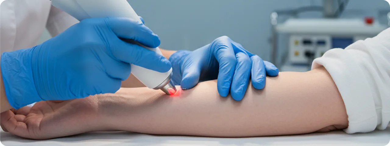
- Laser removal (CO₂ or erbium laser): optimal for small, benign, superficial moles. The laser vaporises the tissue of the lesion layer by layer with minimal damage to the surrounding skin. Advantages: no bleeding, minimal risk of scarring, quick healing (5–10 days). Limitation: it does not allow full histology, so it is used only after dermatoscopy has confirmed that the lesion is benign.
- Radiofrequency removal (Surgitron): high-frequency radio waves cut the tissue without direct contact and without thermal damage. The method combines the advantages of surgery and laser: material can be obtained for histology while tissue trauma is kept minimal. Healing takes 7–10 days. It is widely used for removing raised, papillomatous naevi.
- Cryosurgery (liquid nitrogen): freezing the tissue of the lesion at –196°C. The cells are destroyed by the formation of ice crystals. The method is simple and quick, but it has substantial drawbacks: the depth of the effect cannot be controlled, there is a risk of hypopigmentation, and no material is obtained for histology. It is used mainly for seborrhoeic keratoses and viral warts, and rarely for naevi.
Safety and possible risks
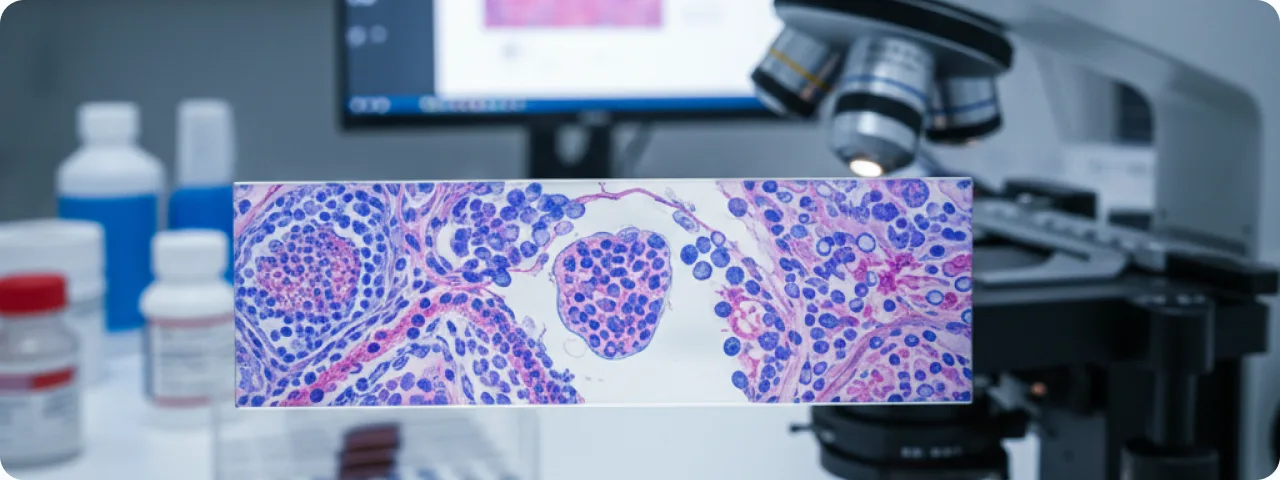
Mole removal is a safe procedure provided the correct protocol is followed and it is performed by a qualified doctor. The main safeguards are: mandatory dermatoscopy before removal to establish the nature of the lesion, local anaesthesia for the patient's comfort, sterility of the instruments and the operating field, and histological examination of the removed material (except for laser removal of lesions confirmed to be benign). Complications are rare and include: a hypertrophic or keloid scar (more often in people with a genetic predisposition), hypo- or hyperpigmentation of the area after healing (usually temporary), wound infection where aftercare advice is not followed, and incomplete removal with recurrence (requiring a repeat procedure). It is important to understand: removing a benign mole does NOT provoke skin cancer — this is a widespread myth with no scientific basis.
Preparing for the procedure
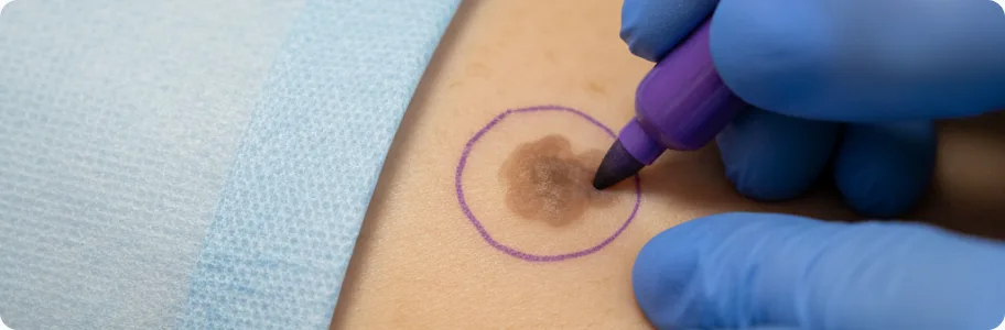
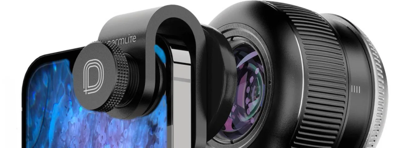
Preparation begins with a dermatological consultation, which includes: the history (when the mole appeared, whether it has changed, any family history of skin cancer), dermatoscopic examination of the lesion with photographs, and a discussion of the method of removal and the expected result. Before the procedure it is recommended: do not apply cosmetics or tanning products to the area for 3 days beforehand; tell the doctor if you take anticoagulants (aspirin, warfarin) — they may need to be stopped temporarily; avoid intensive sun exposure for 2 weeks before the procedure (tanned skin heals less well and carries a higher risk of pigmentation). The procedure itself is usually done as an outpatient and takes 15–30 minutes. Anaesthesia is local infiltration (an injection of lidocaine around the lesion). After removal the doctor gives detailed aftercare instructions: cleaning with an antiseptic, applying a dressing, limiting physical activity and protecting the area from the sun. Sutures (after surgical excision) are removed after 7–14 days depending on the site.
Aftercare and recovery
Proper aftercare is the key to a good cosmetic result and to preventing complications. The first 24–48 hours: keep the wound dry, do not remove the dressing, avoid physical exertion. The first week: clean daily with an antiseptic (chlorhexidine or miramistin), apply a sterile dressing or a special plaster, and avoid steam baths, saunas and swimming pools. Until healing is complete (2–4 weeks): do not pick off the crust — it will come away on its own; protect the area from direct sunlight (SPF 50+ or a dressing); do not apply make-up over the wound. After healing: use scar treatments (silicone sheets, silicone gels) as recommended for 2–3 months, and continue sun protection (SPF 50+) for 6–12 months to prevent hyperpigmentation. A follow-up appointment is arranged 2–4 weeks after removal to assess healing and to give the histology results. If histology shows atypical cells, the doctor will discuss whether a wider excision or additional follow-up is needed. Regular preventive examinations with dermatoscopy are recommended annually to monitor other lesions.
Common questions about mole removal
Does removing a mole hurt? — The procedure is performed under local anaesthesia, so the patient feels no pain during removal. Once the anaesthetic wears off there may be mild discomfort for 1–2 days, relieved by ordinary painkillers. Will there be a scar? — Modern methods minimise scarring. After laser removal of small moles the mark is usually invisible within 3–6 months. Surgical excision leaves a fine linear scar that becomes barely noticeable over time, particularly with proper care. Can moles be removed in summer? — It is better to plan the procedure for autumn or winter, when the sun is least intense. That reduces the risk of pigmentation at the site. Where there are medical indications, however, removal is carried out at any time of year, with sun protection without fail. How much does removal cost? — The cost depends on the method, the size of the lesion, its site and whether histology is needed. The doctor gives the exact cost at the consultation after examining you. Can a mole grow back? — After complete removal, recurrence is unlikely. If a superficial removal was performed (shave biopsy or laser) without taking the deeper layers, residual melanocytes can cause the pigmentation to return — this is not dangerous, but it should be reassessed by a dermatologist.
Related reading
Did not find the answer to your question?
Every case is individual. If you have noticed changes in your skin, or have symptoms that concern you, book a consultation.