Dermatoscopy: how a doctor assesses a skin lesion
What is dermatoscopy?
Dermatoscopy (dermoscopy, epiluminescence microscopy) is a non-invasive method of examining the skin that allows the doctor to see structures invisible to the naked eye. Using a special instrument — the dermatoscope — the doctor magnifies the image 10–20 times and assesses the morphological structures of the epidermis, the dermo-epidermal junction and the superficial dermis. The method entered clinical practice in the 1990s and has since become the gold standard for diagnosing pigmented and non-pigmented skin lesions. Dermatoscopy increases the accuracy of melanoma diagnosis by 30–50% compared with clinical examination by the naked eye. According to a Cochrane meta-analysis, the sensitivity of dermatoscopy for detecting melanoma is 90–95% and its specificity 70–80%. That means the method detects the great majority of malignant lesions while substantially reducing the number of unnecessary surgical procedures. Today dermatoscopy is part of the routine practice of dermatologists, dermato-oncologists and family doctors in most developed countries, and it is an essential element of the assessment of any suspicious skin lesion.
How a dermatoscope works: the principle and the technology
The dermatoscope removes the reflection of light from the surface of the skin, revealing deeper structures to the doctor. There are several types of instrument:
- Contact (immersion) dermatoscopy: the classic method, in which an immersion fluid (oil, gel, alcohol) is applied between the lens and the skin. The fluid reduces refraction of light at the surface of the stratum corneum and makes it transparent. Polarised light is not needed — the doctor sees the structures of the dermo-epidermal junction directly. This method remains the reference standard for the classic dermatoscopic algorithms.
- Polarised (non-contact) dermatoscopy: a more modern approach, using polarised light to remove surface glare without an immersion fluid. It allows better visualisation of vascular structures, crystalline structures and collagen changes. Most modern dermatoscopes are hybrids, with both polarised and non-polarised modes.
- Digital video dermatoscope: an instrument with a built-in high-resolution camera (up to 140× magnification) connected to a computer. Images are captured, stored in a database and compared with the patient's previous images. This allows changes in lesions to be tracked over time — so-called digital monitoring, or mole mapping.
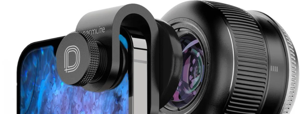
- Mobile dermatoscopy: dermatoscope attachments for smartphones (DermLite, iDerma) democratise the method, making it available to family doctors and for telemedicine consultations. Image quality is inferior to that of dedicated instruments, but sufficient for initial screening and triage. Artificial intelligence is being integrated into mobile applications for automated analysis.
What the doctor sees through the dermatoscope
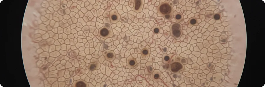
Dermatoscopy opens up a whole world of morphological structures invisible to the naked eye. When examining pigmented lesions, the doctor analyses the pigment network (reticular pattern) — a regular network of brown lines characteristic of benign naevi. Disturbance of its structure — thickening, breaks, irregularity — may indicate malignant transformation. Globules and dots are rounded structures of various sizes and colours (brown, black, blue-grey) corresponding to nests of melanocytes at different depths in the skin. Radial structures (radial streaming and pseudopods) are linear projections at the periphery of a lesion that may indicate active growth of melanoma. Vascular structures are among the most informative criteria: dotted vessels, irregular linear vessels, arborising vessels (typical of basal cell carcinoma) and hairpin vessels (characteristic of keratoacanthoma and squamous cell carcinoma). Regression structures — a blue-white veil, scar-like depigmentation, peppering — indicate partial destruction of the lesion by the immune system.
When dermatoscopy is indicated
Dermatoscopy is indicated in a wide range of clinical situations. Initial assessment of new lesions: any new mole appearing after the age of 30 requires dermatoscopic assessment — new naevi in adulthood are rare and melanoma must be excluded. Monitoring atypical naevi: patients with multiple naevi (more than 50), dysplastic naevi or a family history of melanoma need regular digital dermatoscopy every 6–12 months. Differential diagnosis: dermatoscopy helps to distinguish seborrhoeic keratosis from melanoma, basal cell carcinoma from dermatofibroma, and a haemangioma from a pigmented lesion — cases where clinical examination gives no clear answer. Assessment before removal: before any surgical or laser removal of a lesion, dermatoscopy is essential to establish its nature and to plan the extent of the procedure. Screening of risk groups: people with fair skin (phototype I–II), red hair, a history of sunburn, immunosuppression or previous skin cancer need an annual full skin examination with dermatoscopy. Assessment of non-pigmented lesions: modern dermatoscopy is effective not only for pigmented lesions but also for diagnosing dermatofibromas, angiomas, viral warts, molluscum contagiosum, scabies and other dermatoses.
How to prepare for the examination
Dermatoscopy needs no special preparation and is entirely painless and safe. Duration: examining a single lesion takes 1–2 minutes; a full body examination (total body dermoscopy) takes 20–30 minutes. Before the appointment it is advisable to remove make-up from the areas to be examined. Do not use fake tan for a week beforehand — artificial pigment can distort the dermatoscopic picture. If you monitor your own skin, bring photographs of the lesions that concern you — this helps the doctor to assess change over time. At the appointment the doctor will ask about the family history (melanoma, skin cancer in relatives), your personal history (previous removals of lesions, sunburn) and medication (immunosuppressants, photosensitisers). The examination is carried out at room temperature: the doctor examines each area of the body in turn, paying particular attention to lesions on exposed skin, on the scalp and between the fingers and toes. The findings are recorded in a report describing each lesion according to standardised algorithms (pattern analysis, the 7-point checklist, the ABCD rule). Where necessary the doctor will take digital images for future monitoring.
Digital dermatoscopy and monitoring
Digital dermatoscopy is the evolution of the classic method, combining optical magnification with computerised capture and analysis of images. During a digital dermatoscopy session, every lesion is photographed and linked to an anatomical map of the patient's body — a mole passport is created. At subsequent visits (usually after 3–12 months) the doctor compares the new images with the previous ones using specialised software that automatically detects changes in the size, shape, colour and structure of lesions. Even minimal changes, invisible to the naked eye, are recorded and analysed. Artificial intelligence substantially extends what dermatoscopy can do: deep learning has reached an accuracy comparable to that of experienced dermatologists. Algorithms analyse thousands of image parameters and give the probability that a lesion is malignant. AI does not replace the doctor, however — it is a supporting tool that improves diagnostic accuracy, particularly for less experienced clinicians. The clinical decision always rests with the dermatologist, who takes account of the context: the history, the phototype, how the lesion has changed and other clinical features unavailable to an algorithm. Recommended frequency of monitoring: every 3 months for high-risk patients (multiple atypical naevi, a history of melanoma), every 6–12 months for moderate risk, and annually for the general population over 35.
Related reading

Did not find the answer to your question?
Every case is individual. If you have noticed changes in your skin, or have symptoms that concern you, book a consultation.