Caring for the skin after a lesion is removed
What happens to the skin after removal?
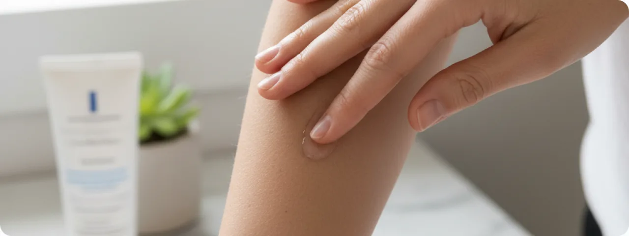
Removing a skin lesion — a mole, papilloma, keratosis, wart or other growth — is controlled injury, after which the body starts its natural process of healing. Whatever the method of removal (laser, radiofrequency, surgery or cryosurgery), the skin goes through successive stages of repair. In the first hours after the procedure a protective crust forms — a natural biological dressing, beneath which the tissue regenerates. The depth and area of the wound depend on the size of the lesion, its site and the method chosen. Superficial lesions (papillomas, keratoses) leave shallow wounds that heal in 7–14 days. Deeper lesions (naevi, dermatofibromas) need 3–4 weeks to heal completely. It is important to understand that the quality of healing depends 50% on proper care at home. Following the doctor's advice substantially reduces the risk of complications — infection, coarse scarring, pigmentary change. Proper care begins immediately after the procedure and continues until the skin has fully recovered.
The stages of wound healing after removal
Healing goes through three successive phases, each of which needs its own care:
- The inflammatory phase (days 1–5): a natural inflammatory reaction develops immediately after removal — redness, swelling and possibly slight pain. This is a normal response. A protective crust of dried lymph and blood forms. During this period it is strictly forbidden to pick off the crust, to wet the wound, to apply make-up or to use a sauna or swimming pool. Clean the wound with an antiseptic (chlorhexidine or miramistin) 2–3 times a day as prescribed.
- The proliferative phase (days 5–21): new tissue forms actively beneath the crust — granulation tissue, then epithelium. The crust gradually separates on its own. The new skin beneath it is pink, thin and sensitive. Continue using the antiseptic and, as prescribed, apply healing preparations (dexpanthenol, products with hyaluronic acid). Protect the area from direct sunlight.
- The remodelling phase (from day 21 to 6–12 months): after the crust has come away, the young skin gradually matures. At first it is pink or red, and in time it takes on a normal colour. Temporary hyper- or hypopigmentation may occur. At this stage sun protection (SPF 50+) is critical in preventing persistent pigmentation. Use scar prevention products as recommended by the doctor.
- Individual differences in healing: the speed and quality of recovery depend on the patient's age, the site of the wound (the face heals faster than the trunk and limbs), general health, chronic disease (diabetes, immunodeficiency), habits (smoking slows regeneration by 30–40%) and a genetic tendency to keloid scarring. Discuss an individual care plan with your doctor, taking these factors into account.
The basic rules of wound care
Proper care is the key factor in successful healing. The first 24–48 hours: keep the wound dry, apply nothing that has not been prescribed, and avoid exertion and overheating. Cleaning the wound: use antiseptic solutions (chlorhexidine 0.05% or miramistin) 2–3 times a day, applied with a cotton bud or a sterile swab. Do not use alcohol-based solutions (iodine, brilliant green) — they cause a chemical burn of the young tissue and slow healing. Do not touch the crust: it will come away by itself in 7–14 days. Removing it early prolongs healing, increases the risk of infection and leads to scarring. Once the crust has come away, apply healing preparations with dexpanthenol or hyaluronic acid in a thin layer twice a day. Sun protection is essential from day one: while the wound is under a crust, cover it with a plaster when going outdoors; once the crust has gone, apply SPF 50+ sunscreen every 2 hours spent in the sun. Active sun protection should continue for at least 6 months, ideally 12. While the wound is healing, avoid swimming pools, saunas, open water, strenuous exercise and alcohol (for the first 3–5 days).
Preventing scars and pigmentary change
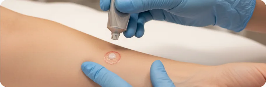
Scar formation is the natural outcome of healing in deeper wounds, but how noticeable it is can be greatly reduced. Silicone products (gels, sheets) are the gold standard for preventing hypertrophic and keloid scars. Begin using them once the wound has closed completely (the crust has gone and the surface has epithelialised) and continue for at least 2–3 months, ideally 6. Silicone gel is applied in a thin layer twice a day; sheets are worn 12–24 hours a day. They work by maintaining optimal hydration and pressure on the scar tissue, which normalises collagen synthesis. Sun protection is a critical element in preventing post-inflammatory hyperpigmentation. Ultraviolet light stimulates the melanocytes in the healing area, which leads to persistent dark patches. Use SPF 50+ every day, even in cloudy weather — UV rays penetrate cloud. Choose products with physical filters (zinc oxide, titanium dioxide) — they irritate sensitive skin less. Scar massage — gentle massage with pressure begins 3–4 weeks after the wound has healed completely. Use circular movements for 5 minutes 2–3 times a day with a moisturiser. Massage improves the elasticity of scar tissue and reduces thickening. If you are prone to keloids, or the wound is in a high-risk area (shoulders, sternum, earlobes), discuss additional preventive measures with your doctor: corticosteroid injections, laser therapy, compression therapy.
When to seek help urgently
Most removals heal without complications when the advice is followed. There are, however, warning symptoms that call for immediate medical attention. Increasing pain after 2–3 days — normal pain gradually subsides; if it increases, or a throbbing pain appears, that may indicate infection. Redness and swelling spreading beyond the wound — local redness in the first days is normal, but if the red area is enlarging and the skin around the wound feels hot, that is a sign of infection. Purulent discharge — yellow-green discharge with an unpleasant smell indicates bacterial infection and requires antibacterial treatment. A temperature above 37.5°C together with local symptoms. Bleeding that does not stop — slight bleeding in the first hours is normal, but prolonged or heavy bleeding requires medical attention. A change in the colour of the wound — black, blue or grey-green areas may indicate tissue necrosis. Do not self-treat: prescribing yourself antibiotics, using home remedies or ignoring warning symptoms can lead to serious complications. Seeking help in good time resolves the problem quickly and prevents complications from developing.
Common questions about aftercare
When can I wash the treated area? — For the first 24–48 hours do not wet the wound at all. After that, wash carefully, avoiding prolonged contact with water and not softening the crust. The area can be washed normally once the crust has come away completely. Swimming pools, saunas and open water — not sooner than 2–3 weeks after the procedure. Can I use make-up over the site? — Decorative make-up (foundation, powder) is not recommended until healing is complete (the crust has gone and the surface has epithelialised). Skincare products only as prescribed. Sunscreen is essential once the crust has come away. How long does full healing take? — It depends on the method and the depth: superficial removal (laser, cryosurgery) — 7–14 days until the crust comes away, 1–2 months until the colour returns to normal. Surgical excision — 10–14 days until the sutures are removed, 3–6 months until the scar has formed. Complete remodelling of the scar takes up to 12 months. Will a mark be left behind? — With proper aftercare the mark is minimal: after laser removal it is usually invisible, or a minimal flat pink spot that fades in 2–4 months; after surgery, a fine linear scar that gradually pales. The result depends considerably on individual healing and on following the advice given. When is a follow-up appointment needed? — The first check is 7–14 days after removal (or on the day the sutures come out). After that, as the doctor recommends — usually at 1, 3 and 6 months. If the lesion was removed because malignancy was suspected, a review after the histology result is essential.
Related reading

Did not find the answer to your question?
Every case is individual. If you have noticed changes in your skin, or have symptoms that concern you, book a consultation.