Basal cell carcinoma: the most common skin cancer, and a curable one
What is basal cell carcinoma?
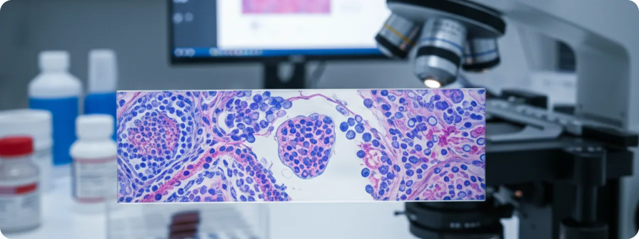
Basal cell carcinoma (basalioma) is the most common malignant tumour of the skin, arising from the basal cells of the epidermis. It accounts for around 75–80% of all cases of skin cancer. More than 3.6 million new cases are diagnosed worldwide each year. Basal cell carcinoma is characterised by slow local growth and metastasises extremely rarely (less than 0.1% of cases), which sets it apart from other malignant tumours. That is why it is sometimes called a "semi-malignant" tumour. Ignoring it is dangerous, however: untreated, it can gradually destroy the surrounding tissue — skin, cartilage, bone — leading to serious disfigurement and loss of function. With early detection and adequate treatment the outlook is extremely favourable: complete cure is achieved in 95–99% of patients. Basal cell carcinoma occurs most often on exposed parts of the body: the face (nose, eyelids, ears), the neck and the scalp — that is, on skin subject to chronic ultraviolet exposure.
Risk factors and causes
Basal cell carcinoma develops through a combination of genetic factors and external exposures. The main risk factors:
- Chronic ultraviolet exposure: the principal risk factor. The cumulative dose of sun exposure over a lifetime plays a key role — sunburn in childhood and adolescence in particular increases the risk 2–3 times. Using sunbeds before the age of 35 raises the risk of basal cell carcinoma by 75%. Outdoor occupations (farmers, builders, fishermen) are an additional factor.
- Skin phototype: people with fair skin (Fitzpatrick phototype I–II), fair or red hair, blue or green eyes and a tendency to burn are at the highest risk. In people with dark skin, basal cell carcinoma is much less common, though not impossible. Genetic syndromes (Gorlin–Goltz syndrome) sharply increase the risk of multiple basal cell carcinomas.
- Age and sex: the risk rises with age — the peak incidence is between 60 and 80, although in recent decades the disease has been appearing in younger people. Men are affected more often than women, in a ratio of about 2:1, which is related to greater sun exposure through work and recreation.
- Immunosuppression and other factors: patients who have had an organ transplant have a 10 times higher risk. Exposure to ionising radiation, chronic ulcers, burn scars, and contact with arsenic and chemical carcinogens also increase the likelihood. A history of previous skin cancer increases the risk of a new basal cell carcinoma by 40% over 5 years.
How to recognise basal cell carcinoma: signs and symptoms
Recognising basal cell carcinoma early is the key to successful treatment. The most common form — nodular basal cell carcinoma — appears as a small, shiny, translucent nodule, pearly or pink, with fine dilated capillaries (telangiectasias) on the surface. In time an ulcer with raised rolled edges may form in the centre — the classic rodent ulcer (ulcus rodens). Superficial basal cell carcinoma looks like a flat reddish or pink patch with a fine pearly rim at the edge, often on the trunk. It can resemble eczema or psoriasis, which delays diagnosis. Morphoeic (sclerodermiform) basal cell carcinoma is the most treacherous type: a flat, yellowish or waxy plaque with indistinct borders, resembling a scar. Its true extent is often considerably greater than what is visible. Pigmented basal cell carcinoma contains melanin and may be brown or black, mimicking melanoma. The main sign common to all types: a wound that does not heal within 3–4 weeks, or a lesion that bleeds intermittently and forms a crust.

Diagnosis: from examination to histology
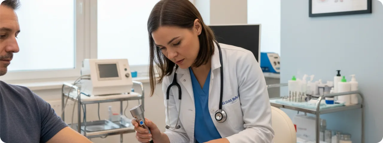
Diagnosis involves several successive steps. Clinical examination: an experienced dermatologist can suspect basal cell carcinoma visually with an accuracy of up to 80%. The site, size, shape and colour are assessed, along with the presence of ulceration and telangiectasia. Dermatoscopy: a non-invasive method that raises diagnostic accuracy to 95–97%. At 10–20× magnification, characteristic structures are revealed: arborising vessels, blue-grey ovoid nests, spoke-wheel areas and leaf-like structures. Dermatoscopy makes it possible to distinguish basal cell carcinoma clearly from benign lesions and from melanoma. Biopsy and histology: the gold standard of diagnosis. An incisional biopsy (part of the lesion) or an excisional one (complete removal) is performed. Histology determines the subtype, which is critical in choosing the approach to treatment. Additional methods: where deep invasion is suspected, high-frequency skin ultrasound (20–75 MHz), MRI or CT is used to assess how far the tumour extends into deep structures. Optical coherence tomography (OCT) is a newer technique for assessing tumour margins non-invasively.
Treatment: current approaches
The choice of treatment depends on the subtype, the size, the site and the risk factors for recurrence. Surgical excision with a 3–5 mm margin remains the gold standard for most basal cell carcinomas: effectiveness is 95–98%, and it allows complete removal to be verified histologically. Mohs micrographic surgery is the most precise method: the tumour is removed layer by layer with 100% of the surgical margin examined under the microscope. It is indicated for recurrent tumours, for lesions in the H zone (nose, eyelids, ears, lips) and for morphoeic forms. Its effectiveness reaches 99%. Cryosurgery with liquid nitrogen is used for small superficial lesions. Photodynamic therapy (PDT) uses a photosensitiser and light to destroy tumour cells — it is optimal for multiple superficial basal cell carcinomas. Topical drug treatment (imiquimod 5%, 5-fluorouracil) is used for superficial tumours where surgery is not possible. Radiotherapy is used in older patients, where there are contraindications to surgery, or as an adjuvant.
Prevention and follow-up after treatment
Prevention rests above all on protection from ultraviolet radiation. Use a sunscreen at SPF 30–50+ every day (even in cloudy weather), applying it 20 minutes before going out and reapplying every 2 hours. Wear protective clothing: a wide-brimmed hat, long sleeves and sunglasses with a UV filter. Avoid the sun between 10:00 and 16:00, when the UV index is at its highest. Give up sunbeds entirely. After successful treatment the patient remains under follow-up: a dermatological examination every 6 months for the first 3 years, then annually. The risk of recurrence is highest in the first 2–3 years and is 5–10% depending on the treatment method and the subtype. The risk of a new basal cell carcinoma within 5 years of the first is 30–50%. A monthly self-examination of the skin using the "new or changing" rule will help to detect a recurrence or a new lesion early. Ask a partner to check areas you cannot see (the back, the scalp). Remember: basal cell carcinoma is a cancer that does not kill, but it can substantially damage quality of life. Timely diagnosis and treatment are the key to complete recovery.
Related reading

Did not find the answer to your question?
Every case is individual. If you have noticed changes in your skin, or have symptoms that concern you, book a consultation.