Melanoma: when to see a doctor
What is melanoma and why is it dangerous?
Melanoma is a malignant tumour of the skin that develops from melanocytes — the cells that produce the pigment melanin. It is the most aggressive form of skin cancer: although it accounts for only 4% of all cutaneous malignancies, melanoma is responsible for 80% of deaths from skin cancer. According to WHO data, more than 325 000 new cases of melanoma are diagnosed worldwide each year, and the incidence is rising by 3–5% annually. The main risk factors are: excessive sun exposure (particularly sunburn in childhood and adolescence), skin phototype I–II (fair skin, red or fair hair, freckles), having more than 50 ordinary naevi or atypical moles, a family history of melanoma, immunosuppression and previous skin cancer. Early diagnosis is critical — when detected at stage I, five-year survival is 99%, whereas at stage IV it is only 15–20%.
Warning signs in a mole (the ABCDE rule)
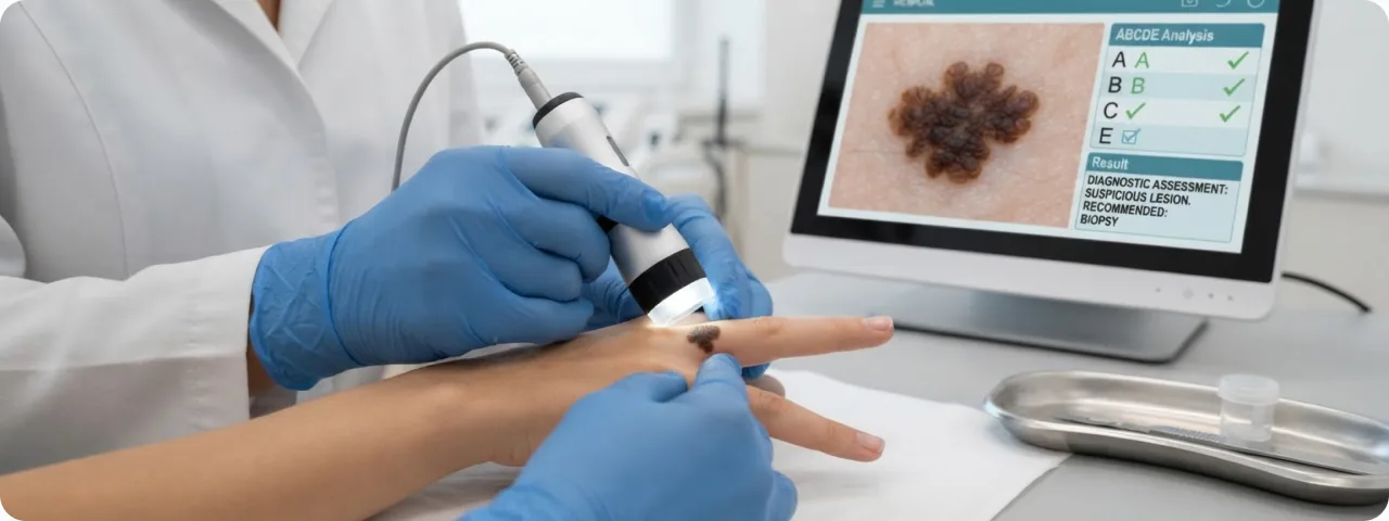
Dermatologists use the ABCDE rule to assess suspicious lesions. Look out for the following:
- Asymmetry: one half of the mole does not match the other in shape, colour or structure. Benign naevi are usually symmetrical. If you draw a line mentally through the centre of the lesion, the two halves should be roughly the same. Asymmetry is one of the earliest and most reliable signs of malignant change and indicates uneven growth of the cells.
- Border: uneven, blurred, notched or scalloped edges. Benign naevi have clear, even borders. In melanoma the edges may look like a map, with projections and indentations. Pseudopods (projections of pigment beyond the main outline) are a particularly worrying sign and indicate invasive growth.
- Color: uneven colouring with several shades of brown, black, red, white or blue within a single lesion. Benign moles usually have one even tone. The appearance of blue-white areas (regression) or black nodules on the surface of a previously evenly coloured mole calls for immediate consultation.
- Diameter and Evolution: a mole larger than 6 mm across (the size of a pencil eraser), or any lesion that changes over time — growing, changing colour or shape, becoming raised, starting to bleed or to itch. Evolution is considered the most important criterion — even a small mole that is changing quickly needs to be examined.
How melanoma is diagnosed
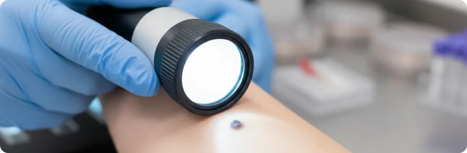
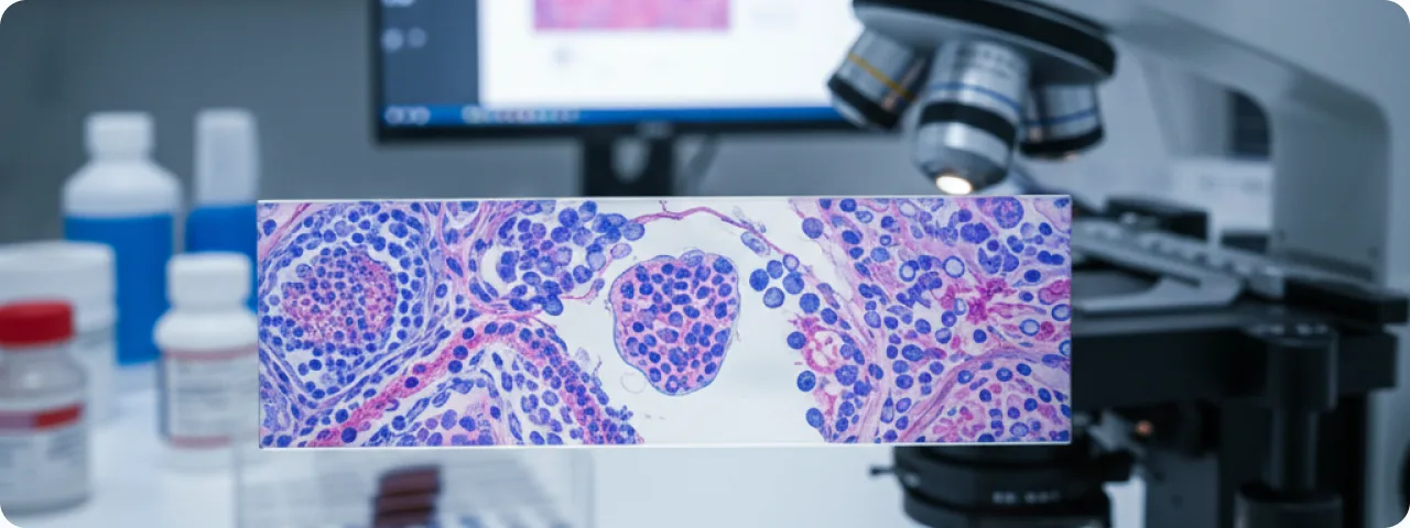
Dermatoscopy (epiluminescence microscopy) is the main non-invasive method and improves diagnostic accuracy by 20–30% compared with examination by the naked eye. The doctor assesses structural patterns: an atypical pigment network, irregular dots and globules, a blue-white veil, regression structures. Digital dermatoscopy with mole mapping allows changes to be tracked at follow-up examinations. Confocal laser microscopy provides non-invasive imaging of skin cells in vivo at a resolution close to histology. Excisional biopsy is the gold standard: the suspicious lesion is removed completely with a 1–3 mm margin for histopathology, with determination of Breslow thickness, Clark level of invasion and mitotic index. Sentinel lymph node biopsy is performed where the tumour is thicker than 0.8 mm, to assess for metastasis.
Current approaches to treating melanoma
Treatment depends on the stage and on the molecular profile of the tumour. Surgical excision is the main treatment for localised melanoma: a margin of 0.5 cm for in situ disease, 1 cm where the thickness is up to 2 mm, and 2 cm where it exceeds 2 mm. Immunotherapy: the checkpoint inhibitors pembrolizumab (anti-PD-1) and ipilimumab (anti-CTLA-4) have revolutionised the treatment of metastatic melanoma, raising five-year survival from 5% to 40–50%. The combination of nivolumab and ipilimumab shows effectiveness of up to 58%. Targeted therapy: where a BRAF V600 mutation is present (40–50% of melanomas), a combination of BRAF and MEK inhibitors (dabrafenib plus trametinib, or vemurafenib plus cobimetinib) produces a response in 70% of patients. Adjuvant therapy after surgery in stage IIB and above reduces the risk of recurrence by 35–40%. Radiotherapy is used for inoperable sites or as palliative treatment for brain metastases.
Prevention and self-examination
Primary prevention: avoid the sun between 10:00 and 16:00, use sunscreen at SPF 30+ every 2 hours (even in cloudy weather), and wear protective clothing and a wide-brimmed hat. Give up sunbeds entirely — WHO classifies artificial UV radiation as a group 1 carcinogen. Examine your skin every month using the ABCDE method: use a mirror to check your back, scalp and the soles of your feet. Photograph your moles to track changes. A preventive dermatological examination with dermatoscopy is recommended annually for everyone, and every 3–6 months for patients with risk factors. Total Body Photography is recommended in atypical mole syndrome or where there is a family history of melanoma. Teach children and adolescents how to protect themselves from the sun — sunburn at a young age substantially increases the risk of melanoma in adult life.
When to seek help immediately
See a dermato-oncologist immediately if a mole is growing rapidly (visible growth over 1–3 months), is changing colour or becoming uneven in colour, or is becoming asymmetrical or irregular in outline. Urgent consultation is needed if a mole bleeds or ulcerates, if satellite nodules appear around an existing naevus, or if the lesion itches or hurts persistently. See a doctor if a new dark mole appears after the age of 30 (new naevi at that age are rare and deserve attention), or if you have a mole that looks different from all the others (the "ugly duckling" sign). Patients with a family history of melanoma or with many atypical naevi should be followed up regularly. Remember: early diagnosis of melanoma saves lives.
Related reading

Did not find the answer to your question?
Every case is individual. If you have noticed changes in your skin, or have symptoms that concern you, book a consultation.