Mohs surgery: the gold standard for removing skin cancer
Contents
What is Mohs surgery and how does it work?
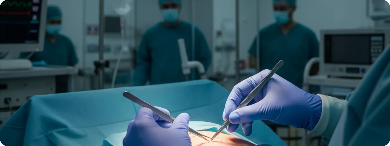
Mohs surgery (Mohs micrographic surgery) is a specialised surgical technique for removing skin cancer that combines staged excision of the tumour with immediate microscopic examination of 100% of the margins of the removed tissue. The method was developed in the 1930s by Dr Frederic Mohs and refined into its modern form in the 1970s. Unlike standard surgical excision, where the removed tissue is sent to a laboratory and the results come back after several days, Mohs surgery involves histological examination during the operation — the surgeon is also the pathologist. The tumour is removed in thin layers: each layer is frozen immediately, cut into sections, stained and examined under the microscope. If cancer cells are found at the margins of a section, the surgeon knows exactly where they are and removes a further layer of tissue only from the affected area. The process is repeated until the margins are clear. This gives a cure rate of 99% for primary basal cell carcinoma and 97% for recurrent tumours — the highest figures of any treatment for skin cancer. Because the method is so precise, the maximum amount of healthy tissue is preserved, which is critical in functionally and cosmetically important areas — the face, ears, eyelids, nose, lips and fingers.
The stages of the procedure
Mohs surgery is performed as an outpatient procedure under local anaesthesia and consists of several clearly defined stages:
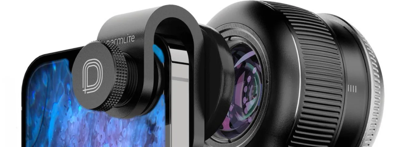
- Stage 1 — preparation and marking: the surgeon examines the tumour, determines its visible borders and marks the area with a special pen. The area is cleaned with an antiseptic and local anaesthesia with lidocaine and adrenaline is given. The patient is awake throughout the procedure. Before starting, the surgeon may use a dermatoscope to define the subclinical borders of the tumour more precisely, which matters particularly in morphoeic and sclerosing types of basal cell carcinoma.
- Stage 2 — excision of the first layer: the surgeon removes a thin layer of tissue (usually 1–2 mm thick) at 45° to the surface of the skin, taking a minimal margin from the visible border of the tumour (1–2 mm, against 4–10 mm in standard excision). The removed specimen is marked with coloured dyes for precise orientation and drawn onto a tissue map — this allows every part of the section to be matched to its anatomical position.
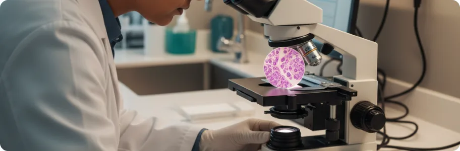
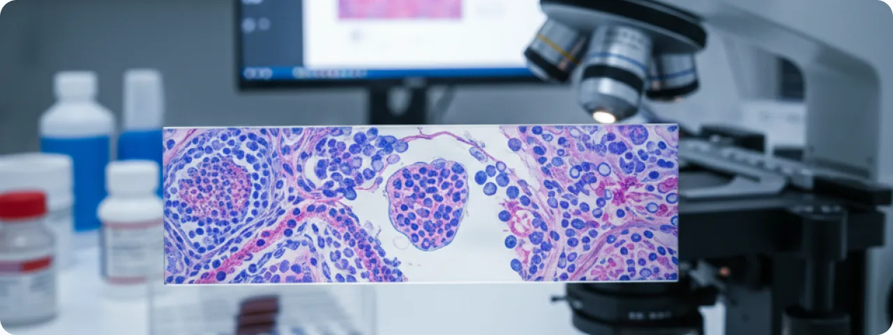
- Stage 3 — processing and microscopy: the removed layer is frozen immediately in a cryostat at –20 to –30°C, cut into horizontal sections 5–8 µm thick, stained with haematoxylin and eosin and examined under the microscope. Unlike standard histology, where less than 1% of the margin is examined, Mohs surgery visualises 100% of the peripheral and deep margins — no area is left unchecked.
- Stage 4 — further excision (if needed): if residual cancer cells are seen under the microscope, the surgeon locates them precisely on the tissue map and removes a further layer only from the affected area, preserving healthy tissue. The cycle repeats until the margins are clear. On average 1–3 stages are needed, although complex cases may require 5 or more. Stage 5 — closing the wound: once clear margins are confirmed, the wound is closed by the most suitable method — primary closure, a local skin flap, a skin graft or healing by secondary intention, depending on the size and site of the defect.
Indications: which tumours are treated with Mohs surgery
Mohs surgery is the treatment of choice for basal cell carcinoma (BCC) and squamous cell carcinoma (SCC) of the skin in particular clinical situations. Location in the "H" zone of the face — the central face, eyelids, eyebrows, periorbital area, nose, lips, chin, ears and temples — is the main indication, because preserving as much tissue as possible here is critical for both function and appearance. Recurrent tumours — those previously treated by other methods (surgery, cryotherapy, radiotherapy) that have returned carry a considerably higher risk of recurring again and require precise margin control. Large tumours (over 2 cm) or those with indistinct clinical borders. Aggressive histological subtypes — morphoeic, infiltrative and micronodular BCC; desmoplastic or acantholytic SCC. Perineural or perivascular invasion — signs that the tumour has grown deeply along nerves or vessels. Tumours in areas where preserving tissue matters functionally — the fingers and toes, the genitals, areas over joints. Immunosuppressed patients — transplant recipients and patients on immunosuppressive therapy have a higher risk of aggressive disease and need the most radical treatment with margin control.
The advantages of Mohs surgery over other methods
Mohs surgery has several key advantages that make it the gold standard in treating skin cancer. The highest cure rate: 99% for primary BCC and 97% for recurrent disease — compared with 92–95% for standard surgical excision and 85–90% for cryotherapy or electrodesiccation. Maximum preservation of healthy tissue: because removal is staged and controlled microscopically, only affected tissue is excised, which reduces the size of the defect by 25–50% compared with standard excision using wide margins. Intraoperative margin control: 100% of the peripheral and deep margins is examined during the operation, unlike standard histology (vertical "bread-loaf" sections), where less than 1% of the margin is seen. A lower risk of recurrence: thanks to complete margin control, the recurrence rate after Mohs surgery is 2–5 times lower than with other methods. Better cosmetic results: a smaller defect means simpler closure, a smaller scar and a better aesthetic result, which matters particularly on the face. An outpatient procedure: it is performed under local anaesthesia with no need for a general anaesthetic or a hospital stay, which lowers both risk and cost. Treatment in a single day: the diagnosis is confirmed and the tumour removed completely at one visit, with no need to return for histology results.
Recovery after Mohs surgery and wound care
Recovery after Mohs surgery is usually quick and predictable. In the first 24–48 hours there may be moderate pain, swelling and slight bleeding — a normal reaction. Pain is controlled with paracetamol; non-steroidal anti-inflammatories (ibuprofen, aspirin) should be avoided for the first 48 hours because of the increased risk of bleeding. The dressing should be kept dry and clean for the first 24 hours; after that it is changed daily, with the wound rinsed with saline and an antibiotic ointment applied (as prescribed). Sutures are usually removed after 5–7 days on the face and after 10–14 days on the trunk and limbs. Complete healing of the wound takes 2–4 weeks, and the scar matures over 6–12 months, during which it gradually pales and becomes less noticeable. For the best cosmetic result it is recommended to protect the scar from the sun for 12 months with SPF 50+ or physical cover. Physical activity should be limited for the first 7–14 days depending on the site: avoid bending, lifting and vigorous exercise, which can put tension on the wound or raise pressure in the operated area. Follow-up includes examinations every 6 months for the first 2 years, then annually for 5 years, since patients with skin cancer have a 30–50% risk of developing a new tumour within 5 years.
Common questions about Mohs surgery
Is Mohs surgery painful? — The procedure is performed under local anaesthesia, so the patient feels no pain during the excision. The anaesthetic injection may cause brief stinging. After the operation the pain is usually moderate and well controlled with ordinary painkillers. Most patients rate the discomfort at 2–3 out of 10. How long does the procedure take? — The duration depends on the size and complexity of the tumour. On average it takes 2–4 hours, including the wait for the microscopy results between stages. Each stage of excision and examination takes roughly 30–45 minutes. We recommend setting aside the whole day. Will there be a scar after Mohs surgery? — Any operation leaves a scar, but because so much tissue is preserved, scars after Mohs surgery are usually smaller than after standard excision. The method of closure (sutures, a flap, a graft) is chosen individually for the best cosmetic result. Does insurance cover Mohs surgery? — Mohs surgery is a recognised medical standard, and most insurers cover it where there are appropriate medical indications. Check the details of your cover with your insurer before the procedure. Can the cancer come back after Mohs surgery? — The risk of recurrence after Mohs surgery is the lowest of any treatment — less than 1% for primary tumours. Patients with skin cancer do, however, have an increased risk of developing new tumours elsewhere, so regular follow-up is essential.
Related reading
Did not find the answer to your question?
Every case is individual. If you have noticed changes in your skin, or have symptoms that concern you, book a consultation.