Nail melanoma: recognising a dangerous streak in time
What is nail melanoma and why is it dangerous?
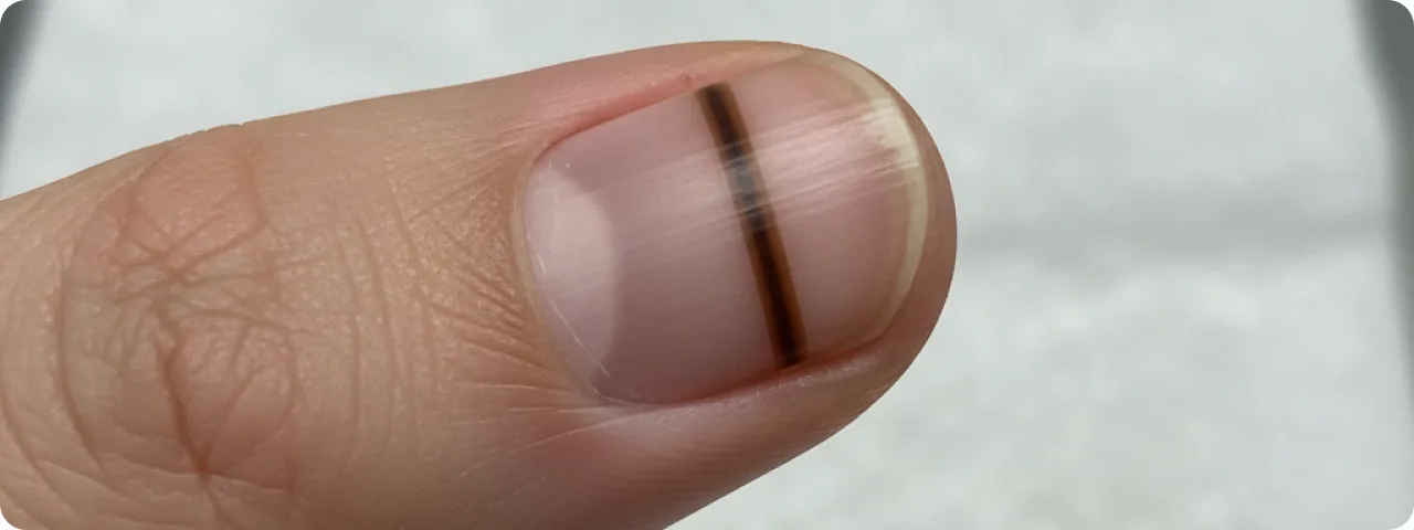
Subungual melanoma is a malignant tumour that develops from the melanocytes of the nail matrix. It accounts for only 1–3% of all melanomas in fair-skinned people, but for up to 15–35% in people with darker skin. The main danger is that the tumour is hidden beneath the nail plate and attracts no attention for a long time. The average interval between the first symptoms and the diagnosis is 2–3 years — considerably longer than for other types of melanoma. That explains why subungual melanoma is diagnosed at later stages and has a poorer five-year survival — around 16–87% depending on the stage at which it is found. The thumb or the great toe is most often affected, because it has the largest area of nail matrix with the greatest number of melanocytes.
Key features: the ABCDEF rule for nail melanoma
Dermatologists have developed a specific ABCDEF rule for assessing suspicious pigmented streaks on the nails, which helps to distinguish benign melanonychia from melanoma.
- A — Age: the peak incidence is between 50 and 70, although nail melanoma can occur at any age. Particular attention should be paid to new pigmented streaks appearing after the age of 50. In children, longitudinal melanonychia is usually benign but should be monitored. Ethnicity matters too: in people with darker skin, melanonychia is considerably more common and is usually a normal variant.
- B — Band: a pigmented band wider than 3 mm, uneven colouring ranging from light brown to black, and blurred or irregular borders are all warning signs. Benign bands are usually narrower, with a uniform colour and clear borders. A band that gradually widens over months is particularly suspicious — this calls for an immediate dermatological consultation and dermatoscopy.
- C — Change: any change in a pigmented band — widening, a change of colour, the appearance of new shades or irregularities — is a warning sign. A stable band that has not changed for years is more likely to be benign. Photographing the nail every 3–6 months makes it possible to track changes objectively and to detect progression in time.
- D, E, F — Digit, Extension, Family: involvement of a single digit (usually the thumb or great toe), spread of pigment onto the nail fold (Hutchinson's sign) and a family history of melanoma are additional risk factors. Hutchinson's sign is one of the strongest predictors of malignancy and calls for urgent biopsy. There is also a pseudo-Hutchinson's sign, however — when dark pigment shows through a translucent cuticle, which is a benign finding.
Diagnosis: from dermatoscopy to biopsy
The first step is dermatoscopy of the nail plate, which allows the colour, width and structure of the pigmented band to be assessed at 10 times magnification. A regular band with parallel lines of uniform colour favours a benign lesion. Irregular lines of varying thickness, interrupted areas and micro-Hutchinson's sign are suspicious. Where warning signs are found, a biopsy of the matrix is performed — the gold standard of diagnosis. Depending on where the pigment lies, a punch, incisional or excisional biopsy is done. It is important that the biopsy covers the whole width of the pigmented band and reaches the matrix. Histopathology determines the Breslow depth of invasion — the key prognostic factor. Immunohistochemical staining (S-100, HMB-45, Melan-A) is used in addition, to confirm the melanocytic origin and to assess the degree of atypia.

Staging and prognostic factors
Once melanoma is confirmed histologically, the stage is determined by the TNM system (AJCC). Breslow tumour thickness remains the most important prognostic factor: at less than 1 mm, five-year survival exceeds 90%, whereas above 4 mm it falls to 45–50%. Surface ulceration, the mitotic index and involvement of the sentinel lymph nodes also affect the outlook. The distance from the growth zone to the bone of the phalanx is minimal, which makes it easier for the tumour to invade deep structures. Satellite metastases, in-transit metastases and involvement of the regional lymph nodes worsen the prognosis. Distant metastases are found most often in the lungs, liver, bones and brain. Early diagnosis is critical: when detected in situ (stage 0), complete cure is possible with surgery.
Treatment: surgery and current methods
The main treatment for subungual melanoma is surgical removal. In the early stages (in situ or with minimal invasion) the digit can be preserved — a wide excision is performed with removal of the whole nail apparatus. In invasive melanoma the standard is amputation at the level of the distal or middle phalanx, although current research shows comparable results with function-preserving surgery for thin tumours. Sentinel lymph node biopsy is recommended where the thickness exceeds 1 mm. For advanced stages, immunotherapy with checkpoint inhibitors (nivolumab, pembrolizumab) and targeted therapy (dabrafenib plus trametinib in BRAF-mutated disease) are used. Adjuvant therapy substantially improves survival where the lymph nodes are involved. Rehabilitation includes fitting a prosthesis for the phalanx and psychological support for the patient.
Prevention and self-examination of the nails
Unlike other forms of melanoma, subungual melanoma is not related to ultraviolet exposure, so sunscreen is not a means of prevention. The key strategy is early detection through regular self-examination. Examine all 20 nails in good light at least once a month. Look for new longitudinal streaks, changes in existing ones, and darkening or destruction of the nail plate without an obvious cause. Photograph any suspicious change for comparison over time. Do not hide dark streaks under polish — that delays diagnosis. Nail injuries (crushing, knocks) can mimic melanoma, but subungual haematomas migrate distally as the nail grows and disappear within 6–9 months. If a dark patch does not go away or is widening, see a dermatologist. People at increased risk (a family history of melanoma, multiple naevi, immunosuppression) should have an annual specialist examination.
Related reading

Did not find the answer to your question?
Every case is individual. If you have noticed changes in your skin, or have symptoms that concern you, book a consultation.